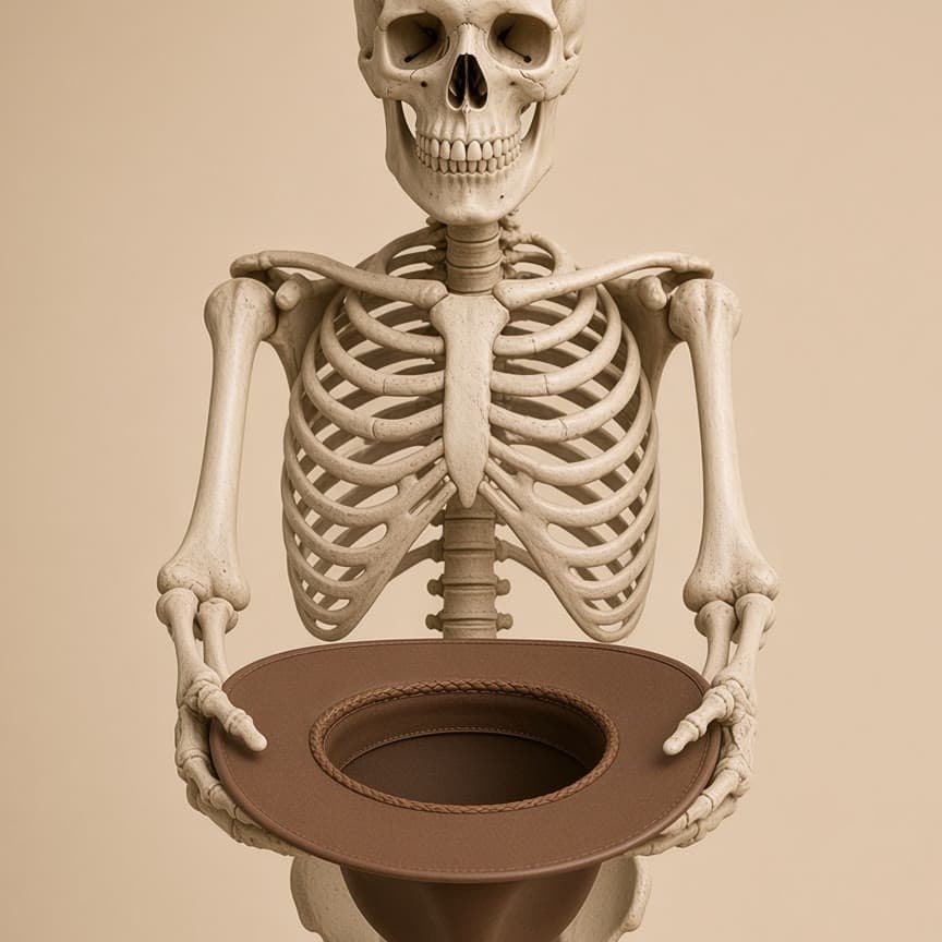
A 420-million-year argument written in tissue
Bone may have been a mineral bank before osteocytes became load sensors.
That is the best-supported interpretation—not a fossil-proven sequence. Early bone was acellular; later osteostracan bone preserves an LCN consistent with local mineral release; modern mechanosensation and osteonal repair were elaborated after that.
Not a straight ladder: vertebrates repeatedly lost, retained, and even reacquired cellular bone.
Three claims to keep separate
Bone came firstEarliest aspidin was acellular dermal bone, with no embedded osteocytes.
Osteocytes came nextFirst clear LCN in osteostracans, with evidence consistent with local mineral release.
Osteons came laterDefinite secondary osteons appear in jawed vertebrates as organized intracortical remodeling.
01 · Nested scales
Cell, network, packet, organ.
The terms describe different scales of organization. Evolution did not invent “the osteon” and then add a cell. It assembled a hierarchy—from cell-scale mineral exchange to organ-scale repair.
02 · The sequence
Four layers of evolutionary reuse.
Each innovation solved a different problem. Later vertebrates stacked the functions together, then tuned or discarded them in different environments.
Layer I · External mineralized armour
The earliest LCN was likely a mineral-access network that inevitably experienced fluid movement under load. Mechanosensation may therefore be an exaptation: a physical consequence of the network, later amplified into biological control. The osteon then became the multicellular repair unit that could act on those local signals.
03 · Deep-time ledger
Not a march from cartilage to bone.
The history includes early mineralized skin, deep origins of endochondral bone, and repeated secondary loss. Sharks are not simply a preserved “pre-bone” stage.
~520 Ma · Conodont claim remains contested
04 · Mechanism explorer
One tissue, four connected problems.
All four causal bridges stay in reach. Pick one, then step the chain from pressure to tissue.
A mineral store is useful only if it can be accessed.
Acellular bone can protect and support. Embedding cells adds microscopic control surfaces throughout the matrix.
Phosphate stored
- Phosphate storedHydroxyapatite locks P and Ca into matrix.
- Osteocyte acidifiesModern cells use proton pumps and proteases.
- Perilacunar matrix dissolvesLacunae/canaliculi locally enlarge.
- Mineral releasedShape and bulk structure remain intact.
Fossil anchor: low-density halos in osteostracan bone are consistent with in-vivo demineralization. They support—not prove—the phosphorus-driver hypothesis.
05 · Natural experiment
Most teleosts discarded the osteocyte—and flourished.
This comparative reversal is the strongest warning against treating modern mammalian mechanosensation as the original, indispensable osteocyte function.
If osteocytes were universally required to sense strain or remodel bone, losing them in most euteleosts should have been crippling. It was not.
06 · Comparative atlas
Evolution preserves functions, not one architecture.
Cellularity, growth rate, and remodeling can vary independently. The combinations reveal what each tissue feature actually does. Pin a row to hold it.
Select a group.
07 · Selection map
No single “reason bone evolved.”
Different pressures explain different layers of the system. Confusing origin with later elaboration produces the usual just-so story. Pin the pressure you want to weigh.
08 · Four-bone comparison
Same tissue family, four opposite strategies.
Ethmoid, otic capsule, skull and talus separate density from turnover, osteocyte number from remodelling, and mechanical importance from mass. “Inner ear bone” here means the otic capsule and bony labyrinth in the petrous temporal; the auditory ossicles belong to the middle ear.
The bone wrapped around the inner ear is among the densest in the body. Close to the hearing organ, it barely renews after birth. The talus is lighter and trabecular, but mechanically dynamic and normally remodels—until its precarious blood supply fails. Ethmoid and skull vault show how thin-sheet and sandwich geometries solve different cranial problems.
Ethmoid
A light, neural-crest-derived separator and olfactory scaffold
- Origin + formation
- Predominantly cranial neural crest; endochondral ossification within the nasal capsule. Its midline mesethmoid and paired ectethmoids fuse in mammals. Human lineage assignment is based mainly on position and comparative/mouse evidence, not direct human fate mapping.
- Architecture
- Pneumatized honeycomb, thin cribriform and orbital plates, scroll-like conchae. The thinnest lamellae are too thin for classic osteons; direct quantitative histology was not found.
- Osteocytes + LCN
- Open gap: no comparable ethmoid lacunar density or LCN metric was found. A sparse sheet-like network is a geometric inference, not a measured result.
- Vessels + loading
- Low intrinsic bone vascularity despite richly supplied nasal mucosa. Very low mechanical load; lightness, separation and airflow matter more than fatigue resistance.
- Mineral + turnover
- Negligible systemic mineral role because mass and inferred turnover are low. No site-specific annual remodelling rate was found.
- Evolution
- Part of the anterior chondrocranium; ectethmoid ossification appeared in multiple tetrapod lineages. Mammals unite several elements into one olfactory bone.
- Clinical consequence
- Failure is usually perforation or fracture, not load fatigue: lamina papyracea can transmit sinus infection to the orbit; cribriform injury can cause CSF leak and anosmia.
- Source
- Chondrocranium / dermatocranium review ↗
Otic capsule
An osteocyte-rich, nearly frozen shell around the labyrinth
- Origin + formation
- Predominantly caudal cranial-base mesoderm around the otic vesicle; endochondral. Human ossification is reported from about 14 centres, beginning around 16 fetal weeks and finishing around 21 weeks.
- Architecture
- Ivory-dense primary, fetal-like endochondral bone with persistent cartilage remnants, hypermineralized patches and an inner-to-outer gradient of remodelling.
- Osteocytes + LCN
- Human inner-layer density is 75,000–90,000 lacunae/mm³; outer layer 50,000–61,000/mm³. Pig inner petrous is about 95,000/mm³ versus mastoid ~28,000 and femoral cortex ~27,000—roughly threefold.
- Vessels + loading
- Extremely low vascularity and essentially no direct mechanical loading. The design target is geometric stability: changing cochlear or semicircular-canal shape can impair hearing or balance.
- Mineral + turnover
- Functionally sequestered from day-to-day calcium/phosphate buffering. High density is not an accessible reservoir when resorption is suppressed.
- Evolution
- Conserved chondrocranial housing for the otic organ; labyrinth shape is phylogenetically informative. Dense, low-turnover petrous bone also preserves ancient DNA unusually well.
- Clinical consequence
- Otosclerosis is the pathological appearance of vascular Haversian foci in normally avascular, non-remodelling bone; it can fix the stapes and cause conductive, then mixed or sensorineural loss.
- Evidence caveat
- OPG diffusion through canaliculi is a proposed remodelling brake, not a complete explanation; human labyrinthitis-ossificans observations imply additional mechanisms.
Skull mosaic
Dermal vault, cartilage-built base, mixed developmental borders
- Origin + formation
- Frontal and most face: neural crest; parietal and much of posterior base: paraxial mesoderm. Vault/face are mainly intramembranous; base is endochondral. Boundaries are mosaic rather than perfectly sharp.
- Architecture
- Vault is a sandwich of outer compact table, diploë with marrow, and inner compact table. Base is massive endochondral bone; face ranges from thin sinus walls to thick masticatory buttresses.
- Osteocytes + LCN
- No single calvarial density value was found. Flat-bone osteocytes are reported rounder than elongated long-bone osteocytes; claims that this makes them more mechanosensitive remain interpretive/model-dependent.
- Osteons + vessels
- Secondary osteons occur in compact tables and thicker facial cortex; diploë remodels on trabecular surfaces. Vascularity is moderate–high through marrow sinusoids, diploic veins, dura and periosteum.
- Loading
- Vault sees low-amplitude radial/tangential stress and impact; facial buttresses carry cyclical mastication. Curvature and sandwich geometry resist bending without long-bone mass.
- Mineral + turnover
- Moderate systemic contribution: more mass and accessible marrow surface than ethmoid or otic capsule, with normal site-dependent remodelling.
- Evolution
- The skull retains two ancient systems: dermal exoskeleton and endochondral chondrocranium. The latter has not been lost in vertebrates; dermal roofing was reduced and reorganized in birds and mammals.
- Clinical consequence
- Calvarial healing is direct intramembranous bone formation rather than the cartilage soft callus typical of long bones. Mouse evidence suggests frontal bone heals better than parietal; human translation is unproven.
- Source
- Cranial-base development review ↗
Talus
A high-load trabecular transmitter with a fragile blood supply
- Origin + formation
- Lateral-plate mesoderm; endochondral cartilage model from about 7 gestational weeks, vascular invasion/cartilage canals at 9–12 weeks, and ossification around the sixth month.
- Architecture
- Thin cortical shell around a predominantly trabecular interior. Trabecular orientation and regional bone volume track locomotor loading; no adult long-bone-style growth plate persists.
- Osteocytes + LCN
- Open gap: no comparable talar lacunar density or site-specific LCN metric was found. A normal lamellar mechanosensing network is expected, but remains unquantified here.
- Osteons + turnover
- Normal cortical osteons where thickness permits, plus active trabecular remodelling. No site-specific annual percentage was verified.
- Vessels
- About 60–70% articular-cartilage coverage and no muscle/tendon attachment leave little vascular entry. An extraosseous neck sling feeds the body retrograde, so displaced neck injury can interrupt supply.
- Loading
- High, cyclic, multidirectional gait load: weight passes tibia → talus → forefoot and calcaneus while the center of pressure shifts from heel strike through push-off.
- Mineral role
- Small systemically but locally dynamic during repair. AVN is a perfusion failure, not a systemic mineral-homeostasis failure.
- Evolution
- The amniote astragalus co-ossified ancestral tarsal elements during ankle reorganization for terrestrial locomotion; exact element homologies remain debated.
- Clinical consequence
- Neck fracture/dislocation can cause avascular necrosis, delayed union and dome collapse, then hindfoot malalignment and post-traumatic ankle/subtalar OA. Revascularization has been reported to take about two years.
- Source
- Talus vascularity / AVN review ↗
Osteocytes do not equal turnover
The otic capsule has by far the highest measured lacunar density here and almost no normal adult remodelling. Osteocytes can preserve a matrix by suppressing replacement, not only trigger it.
Thickness gates the osteon
Classic Haversian remodelling needs vascular canals and enough matrix thickness. Vault tables and talar cortex can support it; the finest ethmoid sheets cannot, while the otic capsule suppresses it despite its thickness.
Blood supply sets the failure mode
Ethmoid fails by perforation, otic capsule by pathological remodelling, skull by suture/defect problems, and talus by vascular interruption followed by necrosis and collapse.
Established comparison
- Vault/face and cranial base use different ossification programs; crest–mesoderm boundaries are mixed.
- Adult otic capsule is avascular, retains primary endochondral bone and normally does not remodel.
- Petrous lacunar density is about three times pig mastoid/femoral cortex in the cited study.
- Talus is endochondral, highly cartilage-covered and supplied retrograde; displaced neck injury threatens viability.
- Calvaria repairs intramembranously; talus relies on perfused trabecular/cortical remodelling.
Open or emerging
- No comparable ethmoid or talus osteocyte-density value was found.
- No separate annual remodelling percentage was verified for ethmoid, calvaria or talus.
- Ethmoid absence of classic osteons is inferred for its thinnest plates, not directly quantified.
- Round calvarial osteocytes being intrinsically more mechanosensitive is model-dependent.
- OPG may help suppress otic-capsule turnover, but it is not a complete explanation.
- Human frontal-versus-parietal healing differences remain unproven; current evidence is from mice.
09 · Honest frontier
What the tissue cannot yet tell us.
Fossils preserve spaces and mineral patterns—not gene expression, signaling molecules, or selective motives. Open a question to hold it.